Visualizing and quantifying tissues and organs at a microscopic level of detail has always been an important tool for biomedical research and daily clinical practice. While many microscopic approaches exist nowadays, only recently there are possibilities to study large pieces of tissue or complete organs at high resolution and in 3 dimensions. However, on the one hand, most techniques still require a lot of tissue processing like fixing, staining or optical clearing to use the (mostly optical) imaging approaches. On the other hand, there are the clinical imaging modalities such as X-ray computed tomography, magnetic resonance and ultrasound imaging which provide great information in clinical practice but have limited resolution or contrast, especially for soft tissues.
Recently, we have shown that using wave-propagation properties of X-rays, rather than only X-ray absorption by tissues, provides a novel way to look at unprocessed tissues and even organs with great contrast and resolution [1,2]. This novel X-ray phase-contrast tomography (X-PCI) requires bright and coherent X-ray beams, which currently can only be produced using synchrotrons, namely large electron accelerators producing bright photon bundles, predominantly for research purposes.
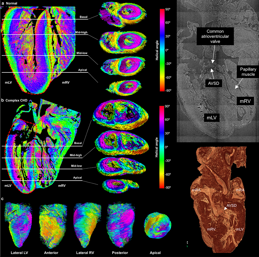
The images below show examples of where this approach can shed light and understanding on microscopic tissue remodelling associated with cardiovascular diseases. Fig 1 shows the damage to the elastic laminas of the aorta in (mouse) Marfan syndrome, of which it is known that patients show gradual damage and enlargement of their aortas, often requiring replacement surgery by the age of 40. Fig 2 shows microstructural details of the heart of a normal human fetus as well as a fetus with congenital heart disease, where the homogeneous unique pattern of cardiac muscle cell orientation is importantly distorted, thus leading to an intrinsic inefficient heart muscle and problems to pump blood around.
References
Garcia-Canadilla P, Dejea H, Bonnin A, Balicevic V, Loncaric S, Zhang C, Butakoff C, AguadoSierra J, Vazquez M, Jackson LH, Stuckey DJ, Rau C, Stampanoni M, Bijnens B & Cook AC 2018, ‘Complex Congenital Heart Disease Associated With Disordered Myocardial Architecture in a Midtrimester Human Fetus’, Circulation-cardiovascular Imaging, 11, 10, e007753.
Lopez-Guimet J, Pena-Perez L, Bradley RS, Garcia-Canadilla P, Disney C, Geng H, Bodey AJ, Withers PJ, Bijnens B, Sherratt MJ & Egea G 2018, ‘MicroCT imaging reveals differential 3D microscale remodelling of the murine aorta in ageing and Marfan syndrome’, Theranostics, 8, 21, 6038 – 6052.
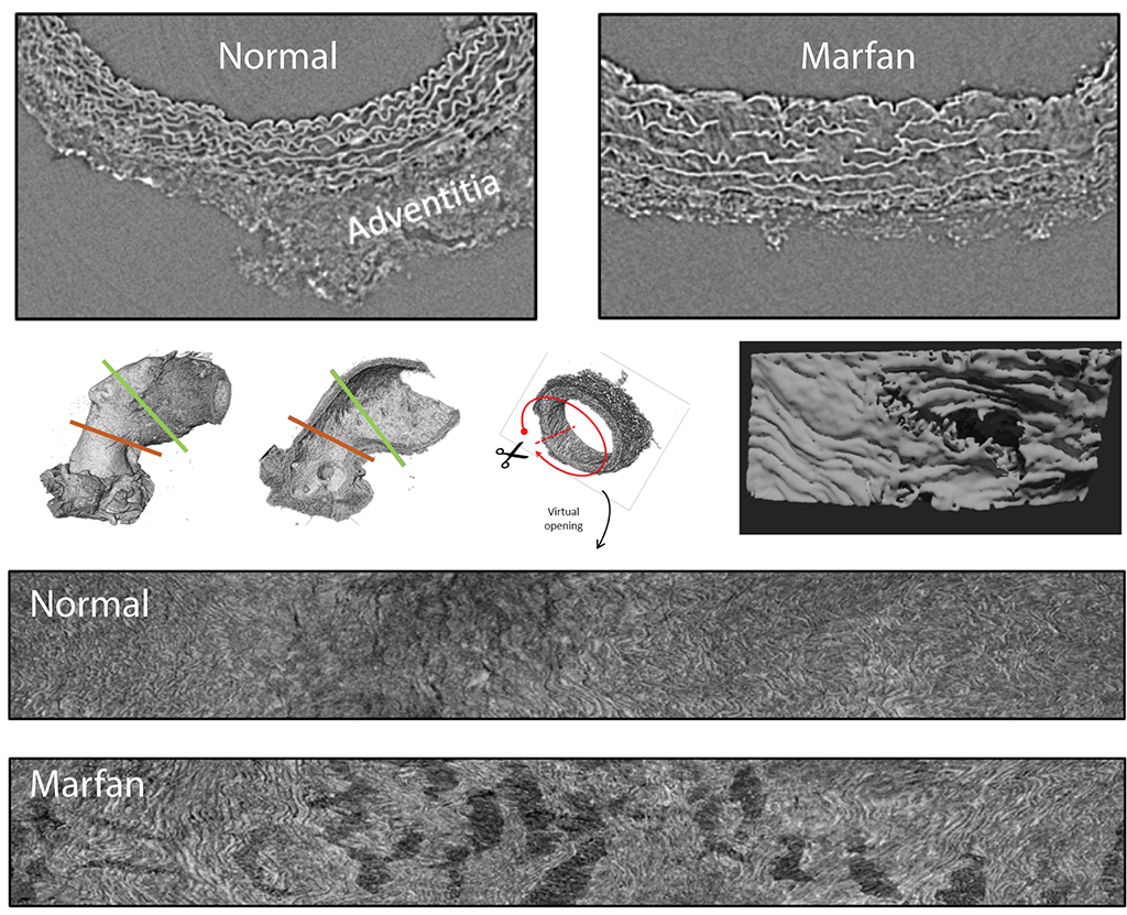
Damage of the elastin laminas in mouse aortas associated by Marfan Syndrome. The top images show local cross-sections of the aorta in adult control and Marfan mice where the interruption of the elastic lamina in Marfan can be clearly observed. Given the 3D nature of the data, a single lamina covering the whole aorta can be isolated and shown (bottom) on a 2D flattened surface to clearly show the fenestrations associated with the disease and which lead to aortic weakening, dilatation and potential rupture.