Holography is known for its ability to produce 3D images (holograms) by recording the amplitude as well as the normally invisible phase of coherent light. In biology though holography is less common, as the most suitable technique, combining sensitivity, resolution and specificity, is fluorescence imaging which is widely used in live cell imaging. It would be fantastic if one could combine fluorescence microscopy with holography, yet fluorescence is incoherent, with a very short pathlength and phase memory.
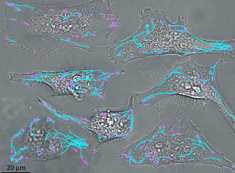
Now ICFO researchers Matz Liebel and Jaime Ortega-Arroyo implemented an interferometry scheme that measures the position-dependent phase change in wide-field fluorescence detection, to enable 3D imaging of individual molecules and nanoparticles with a resolution of 15 nm, over 8 micrometer depth. Teaming up with Hakho Lee at the Massachusetts General Hospital in Boston, they showed how fluorescence holography can track the 3D motion of extracellular vesicles inside live cells.
While fluorescence is preferred for its brightness, Raman response has the advantage of a label-free specific contrast, distinguishing different cellular and tissue contents. Spontaneous Raman scattering is very weak, but luckily can be enhanced dramatically on metal surfaces or in metallic nanogaps: surface enhanced Raman scattering (SERS).
Now, in collaboration with Nicolas Pazos-Perez and Ramon Alvarez-Puebla, of Univ. Rovira i Virgili in Tarragona, Matz Liebel has realized Raman holography for the first time. They synthesized plasmonic superclusters from small nanoparticle building blocks, to generate very strong electric fields in 50 nm cluster size, yielding extremely bright SERS nanoprobes. Next, making the incoherent Raman scattering to “self-interfere”, they achieved 3D holographic imaging, to localize single-SERS-particles in a 3D volume from one single-shot image. Expanding on the fluorescence holography, they now managed to identify and track single SERS nanoparticles inside living cells, in all three dimensions. The SERS signal is bright, photostable and very specific, thus highly suitable for multiplexed singleshot 3D concentration mapping in many scenarios.
Reference
Liebel M, Pazos-Perez N, van Hulst NF & Alvarez-Puebla RA 2020, “Surface-Enhanced Raman Scattering Holography”, Nature NanoTechnology 15 (12) 1005-1011.
Liebel M, Ortega Arroyo J, Sanz Beltrán V, Osmond J, Jo A, Lee H, Quidant R & van Hulst NF 2020, ‘3D tracking of extracellular vesicles by holographic fluorescence imaging’. Sci. Adv. 6 (45), eabc2508
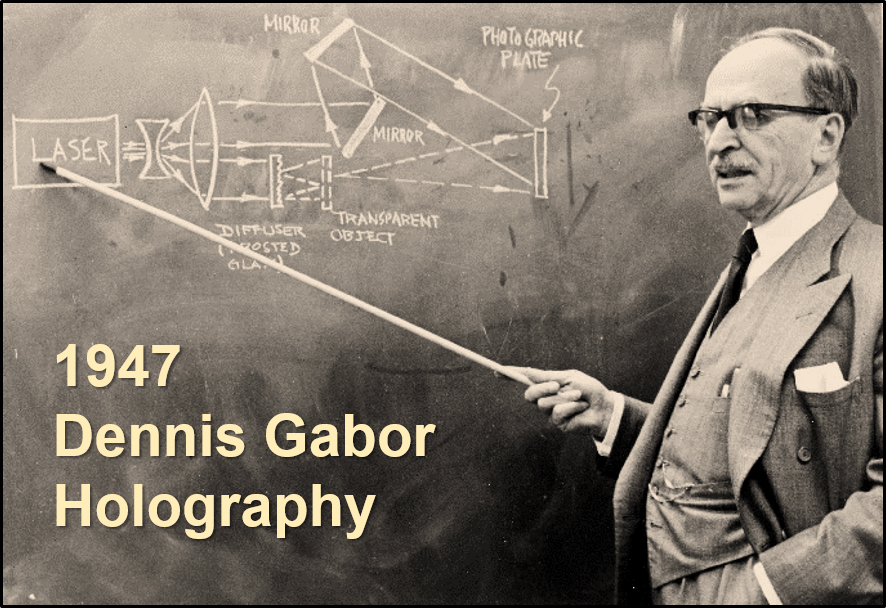
Dennis Gabor (Nobel-Physics 1971) proposes holography: using coherent light illumination and a reference wave, one can record both amplitude and phase, to reconstruct a full 3-Dimensional image of an object. Here using incoherent light, we carry holography into live-cell imaging, with 3D super-resolution and 3D particle tracking.